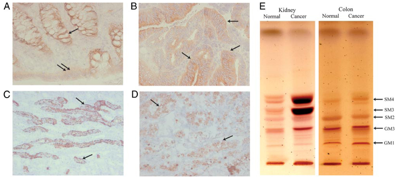
FIGURE 1. Sulfatide can be detected in both normal and cancerous human kidney and colon tissue.
A–D, Frozen samples from human renal and colon tissue were analyzed by IHC. A, Light micrograph showing sulfatide staining in basal crypt epithelium (arrow) and surface epithelium (double arrow) of normal colon (Leica, ×400). B, Colon cancer tissue showing strong intraepithelial (arrows) positivity for sulfatide (Leica, ×200). C, Light micrograph illustrating cortex of normal kidney. Collecting duct epithelium (arrows) shows presence of sulfatide. (Leica, ×200). D, Sulfatide expression (arrows) in renal cell cancer tissue, with positive apoptotic cells (Leica, ×200). E, TLC of acidic glycosphingolipid species. Normal and cancerous tissue of human kidney and colon were analyzed.

