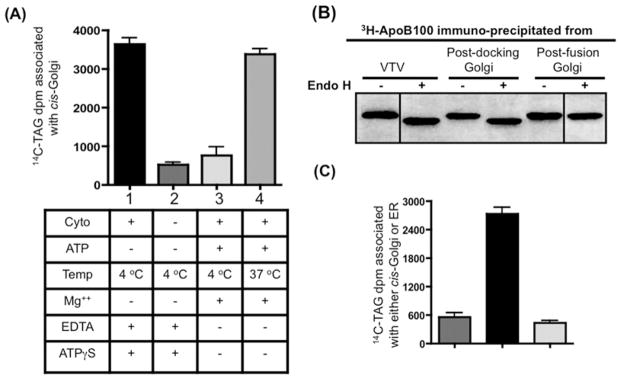
Figure 3. VTVs dock at hepatic cis-Golgi and require cytosol for the docking and fusion process.
(A) VTVs (150 μg of protein) containing [14C]TAG and [3H]apoB100 were incubated with non-radiolabelled cis-Golgi (300 μg of protein) with or without cytosol at either 4 or 37 °C in the presence or absence of Mg2+ and ATP as indicated. After incubation for 30 min, the cis-Golgi fraction was isolated on a sucrose step gradient and collected by aspiration of the 0.86/1.15 M interface, and the amount of [14C]TAG was measured. The Golgi was separated from VTVs and the Golgi-associated [14C]TAG was extracted and levels were measured. Results are means + S.E.M. (n = 4). ATPγS, adenosine 5′-[γ-thio]triphosphate. (B) Exactly the same experiment was carried out as in (A) and the [3H]apoB100 was immunoprecipitated either from the VTVs, post-docking Golgi (A, bar 1) or the post-fusion Golgi (A, bar 4). Immunoprecipitated [3H]apoB100 was incubated with endo H (500 units) for 20 h at 37 °C, separated by SDS/PAGE (5–15 % gel), and autoradiographed. (C) VTVs (150 μg of protein) containing [14C]TAG were incubated with either non-radiolabelled cis-Golgi (300 μg of protein) (left-hand and middle bars) or non-radiolabelled hepatic ER (right-hand bar) at 4 °C in the presence (middle and right-hand bars) or absence (left-hand bar) of cytosol. Mg2+-ATP were excluded in each case. After incubation, the cis-Golgi fraction was isolated and the [14C]TAG levels were determined. Results are means + S.E.M. (n = 4).

