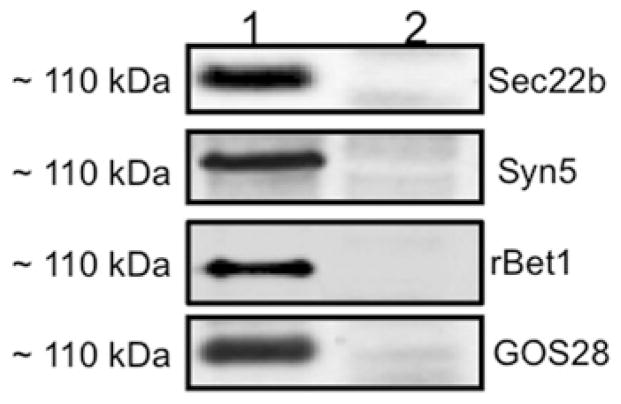
Figure 6. Proteins co-immunoprecipitated with GOS28 in a ~110 kDa complex after VTV–Golgi docking.
A similar VTV–Golgi docking assay was performed as in Figure 5(A), and the VTV–Golgi complexes were isolated. The VTV–Golgi complexes were solubilized in 2 % (v/v) Triton X-100 and incubated with either pre-immune IgG or anti-GOS28 antibodies bound to agarose beads at 4 °C overnight. The beads were washed and either not boiled (lane 1) or boiled (lane 2) in Laemmli buffer. The proteins were separated by SDS/PAGE (5–15 %) and probed with antibodies against the indicated proteins. A single membrane was used, which was sequentially probed with the indicated antibodies after washing. Only protein bands migrated at ~ 110 kDa are shown. Detection was carried out by ECL. The results are representative of four independent trials.

