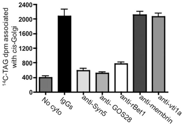
Figure 8. Effect of antibodies against cis-Golgi SNARE proteins on VTV–Golgi fusion.
Hepatic cis-Golgi (300 μg of protein) was incubated with either pre-immune IgG, or antibodies against Syn5, GOS28, rBet1, membrin and vti1a for 1 h at 4 °C. The cis-Golgi membranes were then washed to remove unbound antibody. After antibody treatment, native [14C]TAG-loaded VTVs were added to tubes containing antibody-treated cis-Golgi. The VTV and cis-Golgi were allowed to fuse by incubating them for 30 min at 37 °C with hepatic cytosol (500 μg of protein) and ATP. No cyto represents a negative control where untreated VTVs and Golgi membranes were used without cytosol in the fusion assay. After incubation, the cis-Golgi proteins were isolated on a sucrose step gradient and the Golgi-associated [14C]TAG levels were counted. Results are means + S.E.M. (n = 4).

