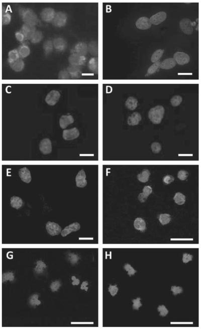
Figure 1.
(A) NCTC2544 cells stably expressing proteinase-activated receptor 2 (PAR2) (clone G) incubated with FITC-labelled SAM11 showed cytoplasmic staining whereas native NCTC2544 cells did not. (B) Clone G cells showed no staining if SAM11 was preabsorbed with its immunising peptide (C) or if incubated with FITC-labelled IgG2a isotype control antibody (D). PAR4-expressing NCTC2544 cells also did not stain when incubated with FITC-labelled SAM11 (E). CD14+ cells from a patient with rheumatoid arthritis (RA) during a fl are (55% surface PAR2 expression) incubated with FITC-labelled SAM11 showed cytoplasmic staining (F) whereas these cells from a healthy control (G) and a patient with osteoarthritis (OA) (H) did not. Nuclei stained blue with 4′,6-diamidino-2-phenylindole. Scale bar=20 μm.

