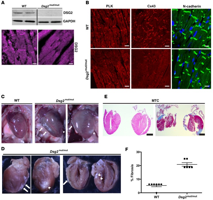
Figure 2. Biventricular fibrosis and abnormal distribution of junctional proteins in Dsg2mut/mut mice at 16 weeks of age.
(A) Western immunoblots and immunolabeled myocardium probed for desmoglein-2 (DSG2) showed a complete absence of protein and junctional distribution of DSG2 in Dsg2mut/mut mice compared with WT mice. Scale bar: 10 μm. (B) Representative images from myocardium immunolabeled for plakoglobin (PLK), connexin43 (Cx43), and N-cadherin. Scale bar: 20 μm. (C and D) Gross pathology shows right ventricle (white arrows) and left ventricle (white arrowhead) epicardial and endocardial scars. (E) Masson’s trichrome–immunostained (MTC-immunostained) myocardium from Dsg2mut/mut mice shows extensive epicardial and endocardial scarring compared with WT controls. Scale bar: 1 mm. Images are representative of n = 7 and n = 6 for WT and Dsg2mut/mut mice, respectively. (F) Percentage fibrosis presented as mean ± SEM. P < 0.05 for WT vs. Dsg2mut/mut using 2-tailed t test with equal variance.

