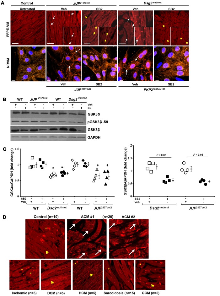
Figure 6. GSK3β localization is uniquely abnormal in ACM, and SB216763 normalizes it.
(A) Representative images of formalin-fixed, paraffin-embedded ventricular myocardia (FFPE-VM) (scale bar: 20 μm) from JUP2157del2 and Dsg2mut/mut mice and neonatal rat ventricular myocytes (NRVM) (scale bar: 10 μm) expressing JUP2157del2 and PKP21851del123 transgenes immunolabeled for anti-GSK3β. Images are representative of n = 4/genotype/treatment (DAPI, blue; GSK3β, red; white arrows, ID localization of GSK3β; yellow arrows, absence of ID localization of GSK3β). (B) Western blots probed for GSK3α, GSK3β, and phosphorylated GSK3β (pGSK3β-S9) from WT, Dsg2mut/mut, and JUP2157del2 mice. (C) Quantitative GSK3β and GSK3α protein levels from WT, Dsg2mut/mut, and JUP2157del2 mice, normalized to GAPDH. Mean ± SEM, n = 4/genotype/treatment. P < 0.05 for SB2-treated mice vs. Veh-treated mice using 2-tailed paired t test. *Dsg2mut/mut vs. WT; ‡JUP2157del2 vs. WT. (D) GSK3β-immunolabeled patient biopsies. Representative images taken from endomyocardial biopsy samples show GSK3β distribution at IDs (white arrows) in all patients with ACM (20 of 20) differ from control hearts (n = 10) and patients diagnosed with sarcoidosis (n = 15), giant cell myocarditis (GCM, n = 5), and end-stage ischemic, dilated cardiomyopathy (DCM), and hypertrophic cardiomyopathy (HCM, n = 5 for each) (yellow asterisks, punctate cytosolic pools of GSK3β; yellow arrows, absence of GSK3β signal at IDs).

