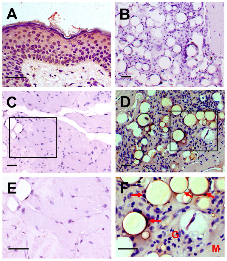
Figure 2.
Histologic sections of muscle stained with antibody to CYP27B1 (25-OH vitamin D 1-α-hydroxylase). (A) Positive control (normal human skin biopsy), (B) Negative control (no CYP27B1 antibody, patient muscle), (C, E) Normal muscle (an unrelated patient), (D, F) Patient muscle (M) with strong CYP27B1 expression (brown) in the histiocytes surrounding presumed PMMA globules (arrows) in the inflammatory reaction (G). Scale bars = 50 μm.

