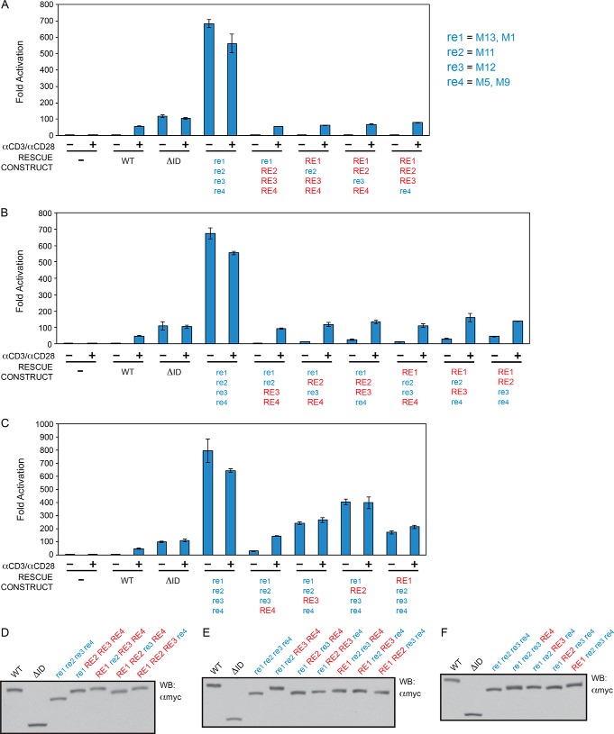
FIGURE 8.
RE1, RE2, RE3, and RE4 cooperatively repress CARD11 signaling to NF-κB. A–C, Jurkat T cells in which CARD11 was stably knocked down (KD-CARD11) were transfected with CSK-LacZ and Igκ2-IFN-LUC in the presence of expression vectors for the indicated Myc-tagged CARD11 variants and stimulated with anti-CD3/anti-CD28 cross-linking for 5 h as indicated. A two-tailed unpaired Student's t test with unequal variance resulted in the following p values in comparison with wild-type CARD11 under unstimulated conditions: re1 re2 re3 re4, p = 0.0004; re1 RE2 RE3 RE4, p = 0.397; RE1 re2 RE3 RE4, p = 0.054; RE1 RE2 re3 RE4, p = 0.0016; RE1 RE2 RE3 re4, p = 0.0054; re1 re2 RE3 RE4, p = 0.0397; re1 RE2 re3 RE4, p = 0.0008; re1 RE2 RE3 re4, p = 0.011; RE1 re2 re3 RE4, p = 0.016; RE1 re2 RE3 re4, p = 0.005; RE1 RE2 re3 re4, p = 0.0012; re1 re2 re3 RE4, p = 0.005; re1 re2 RE3 re4, p = 0.0007; re1 RE2 re3 re4, p = 0.001; RE1 re2 re3 re4, p = 0.0016. D–F, HEK293T cells were transfected with the same amounts of each expression vector used in A–C, and lysates were probed by Western blot using anti-Myc primary antibody to indicate the relative expression level of each variant. β-Galactosidase activity, driven by CSK-LacZ, was used to calculate equivalent amounts of lysate for Western analysis. For ease of representation, the presence of a mutated RE in a construct is indicated by blue lowercase letters (e.g. re1), whereas the presence of a wild-type RE is indicated by red capital letters (e.g. RE1).

