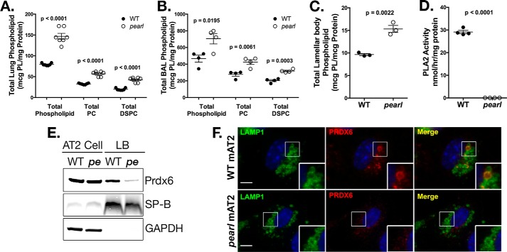
FIGURE 1.
Disrupted lamellar body phospholipid content in pearl mice is associated with loss of lamellar body PRDX6. A, total phospholipid, total PC, andtotal DSPC measured in lung tissue homogenate (n = 6 mice per genotype, mean ± S.E.). B, total phospholipid, total PC, and total DSPC measured in bronchoalveolar lavage fluid from wild type (WT) and pearl mice (n = 4 mice per genotype, mean ± S.E.). C, total phospholipid content measured in lamellar body fractions from WT and pearl mice (n = 3 samples per genotype, each prepared from three mice, mean ± S.E.). D, phospholipase A2 activity measured in lamellar body fractions isolated from WT and pearl mice (n = 4 samples from 2 to 3 mice of each strain, mean ± S.E.). E, representative immunoblots of PRDX6, SP-B, and GAPDH using lamellar body fractions (LB; 10 μg of protein) and alveolar type 2 cell lysate (AT2; 30 μg of protein) from wild type (WT) and pearl (pe) mice. F, immunolocalization of PRDX6 in WT and pearl isolated alveolar type 2 cells (mAT2) after depletion of cytosolic constituents, using LAMP1 immunostaining to identify lamellar bodies (representative of two experiments; bars, 5 μm).

