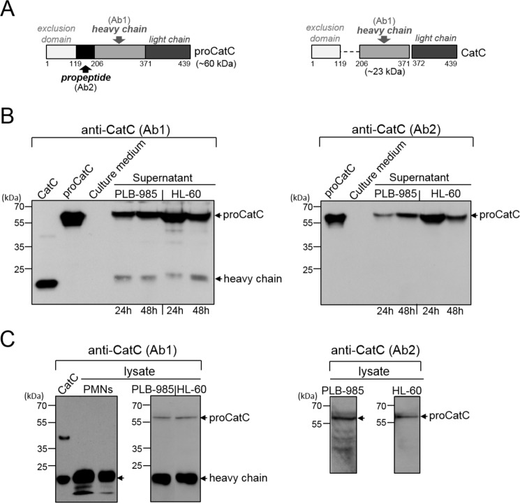
FIGURE 1.
CatC in PLB-985 and HL-60 cells. A, structures of proCatC (left) and mature CatC (right). Arrows indicate the recognition domains of antiCatC antibodies (Ab1, Ab2). B, Western blot analysis of 30-fold concentrated PLB-985 and HL-60 cell supernatants (50 μg of protein/lane) using anti-CatC antibodies Ab1 (left) and Ab2 (right). The cells were cultured for 24 h or 48 h. C, Western blot analysis of PLB-985 cell lysates (50 and 200 μg of protein/lane) and HL-60 cell lysates (50 and 200 μg of protein/lane) using anti-CatC antibodies Ab1 (left) and Ab2 (right). Similar results were obtained in three independent experiments. Recombinant CatC, proCatC, and neutrophil lysates (50 and 20 μg of protein/lane) were used as controls. PMNs, polymorphonuclear neutrophils.

