FIGURE 2.
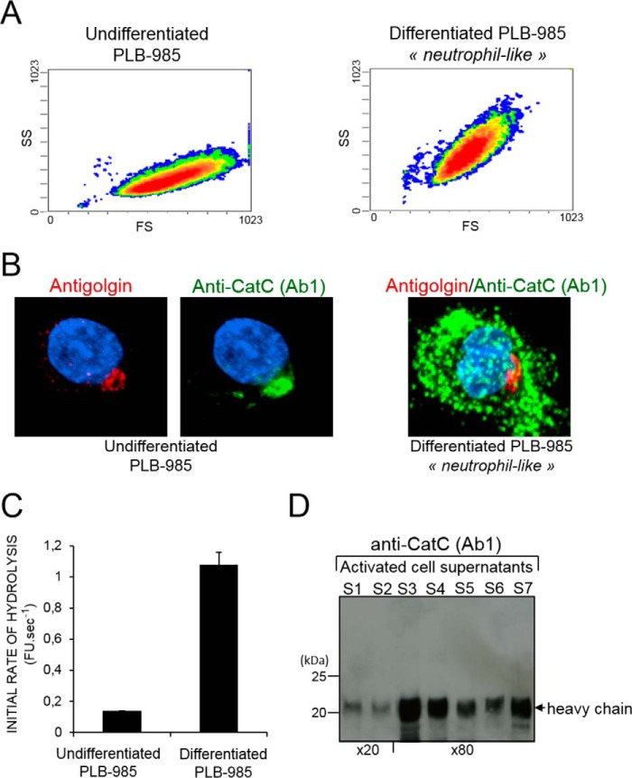
Intracellular localization and secretion of mature CatC by activated cells. A, sideward (SS) versus forward (FS) scatter plot of undifferentiated (left panel) and differentiated PLB-985 cells (right). B, confocal microscopy of undifferentiated (left) and differentiated (right) PLB-985 cells immunostained with anti-CatC (Ab1) antibodies (green) and anti-golgin-84 antibodies (red) showing the initial localization of CatC in the Golgi of immature cells and its distribution throughout the cell after differentiation. C, CatC activity in cell-free supernatants of undifferentiated and differentiated PLB-985 cells after treatment with the calcium ionophore A23187. FU, fluorescence unit. D, immunoblot analysis of 20- or 80-fold concentrated supernatants of neutrophils (S1 to S7) after their activation with A23187 using the anti-CatC antibody Ab1. Similar results were found in three independent experiments.

