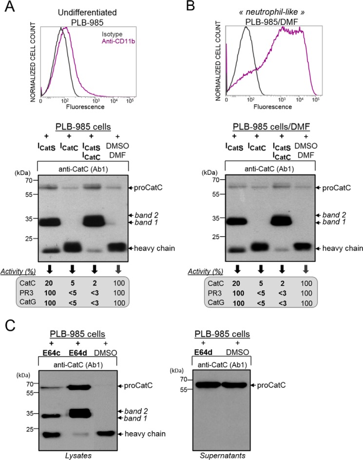
FIGURE 6.
Processing of proCatC in PLB-985 cells cultured in the presence or absence of synthetic inhibitors. A, cell surface CD11b expression was analyzed by flow cytometry. Undifferentiated PLB-985 were cultured for 1 week in the presence of ICatS (10 μm), ICatC (2 μm), or after adding the DMSO/DMF containing buffer alone. Cell lysates were analyzed by Western blotting using anti-CatC antibody (Ab1). The percentage of CatC, PR3, and CatG activity was determined using the respective selective substrates for each protease and is given in the box. B, PLB-985 cells were differentiated into neutrophil-like in the presence of DMF and with or without ICatS (10 μm) or ICatC (2 μm) or DMSO/DMF. Cell lysates were analyzed by Western blotting using anti-CatC antibody (Ab1). The % of CatC, PR3, and CatG activity measured using their respective selective substrates were shown in the box. Cell surface CD11b expression was analyzed by flow cytometry as in A. C, undifferentiated PLB-985 cells cultured in the presence of E64c (100 μm), E64d (100 μm), or DMSO alone for 48 h were lysed. Cell lysates and supernatants of PLB-985 cells were analyzed by Western blotting using anti-CatC antibody Ab1 (left and right panels, respectively). Similar results were observed in five independent experiments.

