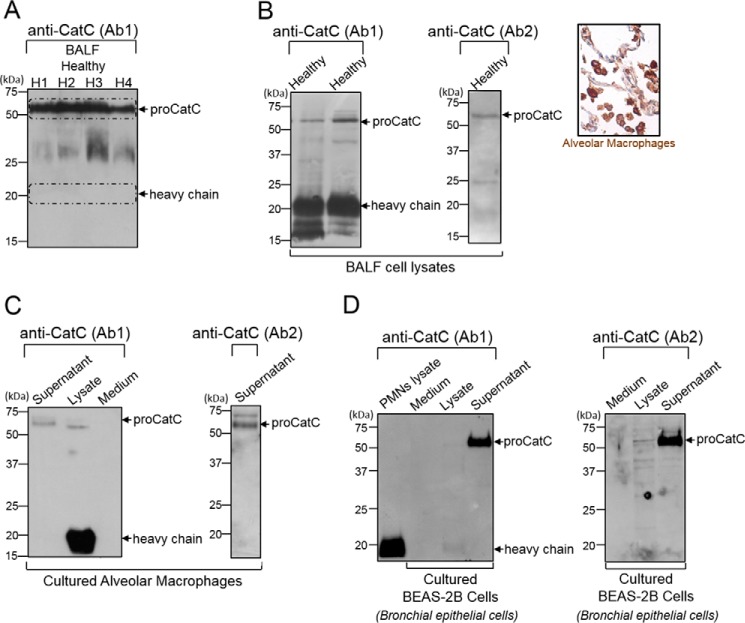
FIGURE 9.
CatC in BALF from healthy subjects and in BALF cell lysates, alveolar macrophages, and BEAS-2B bronchial epithelial cells. A, anti-CatC (Ab1) immunoblots of 30-fold concentrated BALF (H1 to H4) from healthy subjects. Shown are immunoblots with anti-CatC antibodies Ab1 and Ab2 of BALF cell lysates (inset, immunostaining of healthy human lung tissue with Anti-CatC (Ab1) showing the CatC in the cytoplasm of alveolar macrophages) (B), cultured alveolar macrophage lysates and supernatants (C), and cultured BEAS-2B cell lysates and supernatants (D). PMNs, polymorphonuclear neutrophils. Similar results were observed in three independent experiments.

