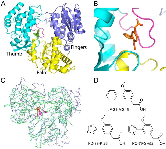
FIGURE 1.
Crystal structure of the DENV-3 polymerase bound to JF-31-MG46. A, the structure of the DENV-3 polymerase is shown as a cartoon, with the palm, fingers, and thumb subdomains colored yellow, blue, and cyan, respectively, and the priming loop colored magenta. The compound, JF-31-MG46, is shown as orange sticks. The difference density, where the compound is removed from the final round of refinement is shown as a green mesh contoured at 3 σ. B, alternative view, colored as in A showing the compound binds between the priming loop and the thumb and palm subdomains. C, alignment of the DENV-3 polymerase shown as blue ribbons with the hepatitis C virus polymerase (Protein Data Bank code 3HKY) bound to a palm site I inhibitor, shown as green ribbons. The compounds bound to the DENV and hepatitis C virus RdRps are shown as sticks colored orange and magenta, respectively. D, the chemical structures of the compounds used in these experiments.

