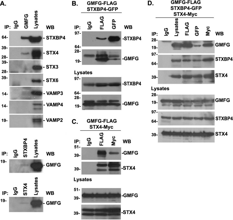
FIGURE 8.
GMFG interacts with STXBP4 and STX4 in human monocytes. A, endogenous GMFG was immunoprecipitated (IP) from THP-1 cells, and precipitates were analyzed by Western blotting (WB) using anti-STXBP4, anti-STX, or anti-VAMP antibodies as indicated (top panel). Endogenous STXBP4 and STX4 were immunoprecipitated from THP-1 cells, and precipitates were analyzed by Western blotting using anti-GMFG antibody (bottom panel). Samples of the total lysate are shown in the right lane, and nonspecific mouse IgG was used as a negative control. B and C, HEK-293T cells were cotransfected with FLAG-tagged GMFG and GFP-tagged STXBP4 (B) or FLAG-tagged GMFG and Myc-tagged STX4 (C). Forty-eight h after transfection, cells were harvested and immunoprecipitated with anti-FLAG, anti-GFP, or anti-Myc antibodies, and precipitates were analyzed by Western blotting with anti-GMFG, anti-STXBP4, or anti-STX4 antibodies. Samples of the total lysates are shown in the bottom panel; each sample corresponds to 5% of the cell lysate used in each immunoprecipitation. D, HEK-293T cells were cotransfected with FLAG-tagged GMFG, GFP-tagged STXBP4, and Myc-tagged STX4. Forty-eight h after transfection, cells were harvested and immunoprecipitated with anti-FLAG, anti-GFP, or anti-Myc antibodies and analyzed by Western blotting with anti-GMFG, anti-STXBP4, or anti-STX4 antibodies. Samples of the total lysates are shown in the bottom panel; each sample corresponds to 5% of the cell lysate used in each immunoprecipitation.

