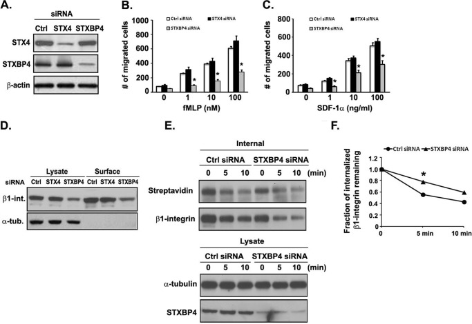
FIGURE 9.
Knockdown of STXBP4 inhibits human monocyte migration and is correlated with inefficient β1-integrin recycling to the cell surface. A, knockdown efficiency of STXBP4 and STX4 siRNA in THP-1 cells. Cells were transfected with a control negative siRNA (Ctrl), STX4 siRNA, or STXBP4 siRNA, and lysates were examined by Western blotting. β-Actin was used as a loading control. B and C, Transwell migration assays were performed on 5-μm pore filters coated with 10 μg/ml FN in THP-1 cells transfected with control negative siRNA (Ctrl siRNA), STXBP4 siRNA, or STX4 siRNA in response to vehicle alone (0.1% BSA) or increasing concentrations of fMLP (1–100 nm) or SDF-1α (1–100 ng/ml). The number of migrated cells was quantitated after 3 h, as described under “Experimental Procedures.” Data represent the mean ± S.D. (error bars) of at least three independent experiments. *, p < 0.05 compared with control siRNA-transfected cells. D, THP-1 cells transfected with the indicated siRNAs were surface-labeled with 0.5 mg/ml sulfo-NHS-SS-biotin at 4 °C for 1 h. Surface-biotinylated proteins were then isolated using streptavidin beads, followed by Western blotting analysis of β1-integrin. α-Tubulin was used as a loading control. E, biotinylation-based assay of β1-integrin recycling. THP-1 cells transfected with the indicated siRNAs were biotinylated and allowed to internalize surface proteins for 30 min at 37 °C. Surface biotin was removed by reduction (glutathione solution), and internalized proteins were chased at 37 °C for the indicated time periods to allow recycling back to the surface. Surface biotin was reduced again, and cells were lysed. Top, biotinylated cell surface proteins remaining inside the cells were immunoprecipitated using an anti-β1-integrin antibody and subsequently detected by Western blotting analysis using streptavidin-HRP or β1-integrin antibody. Bottom, samples of the total lysates; each sample corresponds to 5% of the cell lysate used in each immunoprecipitation. Shown is a representative Western blot of one experiment (n = 3). α-Tubulin was used as a loading control. F, quantification of β1-integrin recycling determined by densitometry signal as in E. The fraction of internalized β1-integrin remaining was quantified from the signal intensity of internalized protein at each time point relative to control siRNA-transfected cells that had not been placed at 37 °C after the first surface reduction.

