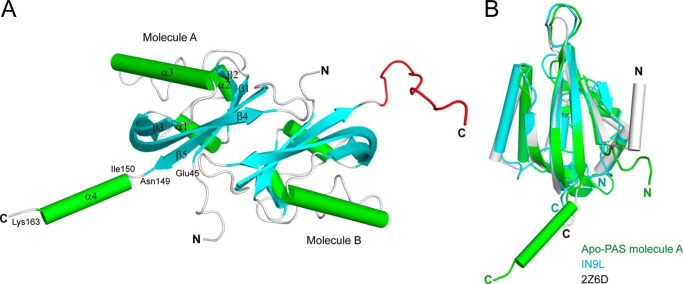
FIGURE 1.
Overall structure of apo-PAS1. A, ribbon representation of the structure of TodS PAS1 in an asymmetric configuration. There are two PAS1 molecules in the asymmetric unit. The β-sheets and α-helices are shown in cyan and green, respectively. The C-terminal disordered region in molecule B corresponding to α4 in molecule A is displayed in red. B, apo-PAS1 (green) superimposed onto the LOV/PAS domain of the C. reinhardtii photoreceptor (Protein Data Bank code 1N9L; cyan) and the LOV1 domain of the Arabidopsis blue light receptor protein phototropin-2 (Protein Data Bank code 2Z6D; gray).

