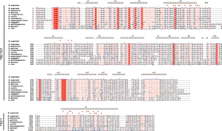
FIGURE 4.
Sequence alignment of TSC1 and TBC1D7 from different organisms. Conserved residues are boxed in red and similar residues are highlighted in red. Residues are framed in blue if more than 70% of its residues are similar according to physicochemical properties. For TBC1D7, helices are marked with squiggles, turns with TT letters, and interface residues with red stars. For the two TSC1 helices interacting with TBC1D7, red stars in the upper row represent TSC1-A residues involved in TBC1D7 interaction, red hashtags in the lower row indicate TSC1-B residues interacting with TBC1D7. For counting convenience, small black dots are added in every 10 residues on top of sequences.

