FIGURE 6.
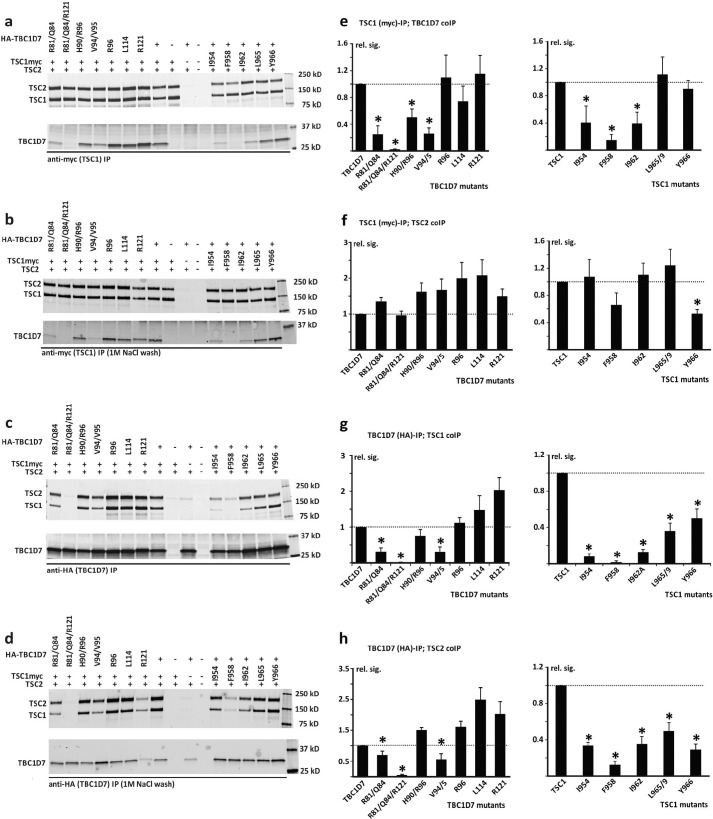
IP analysis of the TSC1-TBC1D7 interaction. C-terminal myc-tagged wild-type TSC1, N-terminal HA-tagged wild-type TBC1D7, or the TSC1 and TBC1D7 variants were coexpressed with TSC2 in HEK 293T cells. TSC complexes were isolated by IP, either with anti-myc affinity beads (specific for exogenous TSC1) or anti-HA affinity beads (specific for exogenous TBC1D7). Four separate transfection experiments were performed. In 3 experiments, IPs were washed 4 times with 20 volumes of lysis buffer. All 3 experiments gave very similar results. In a subsequent experiment, IPs were washed 4 times with lysis buffer and twice with lysis buffer containing 1 m NaCl. a and b, immunoblots showing co-IP of TSC2 and the TBC1D7 variants with immunoprecipitated TSC1. Immunoprecipitates were either washed with lysis buffer only (a), or with high salt buffer (b). c and d, immunoblots showing co-IP of TSC2 and TSC1 with immunoprecipitated TBC1D7. Immunoprecipitates were washed with lysis buffer only (c) or high salt lysis buffer (d), as in A and B. e-h, quantification of the signals on the immunoblots from 3 separate co-IP experiments (no high salt washes). The relative signals of the co-immunoprecipitated proteins are shown relative to the wild-type control, after adjusting for the signal of the immunoprecipitated protein (either TBC1D7 or TSC1). Error bars correspond to the mean ± S.E.; values significantly reduced compared with the wild-type controls (p < 0.05 Student's paired t test) are indicated with an asterisk. e, co-IP of TBC1D7 with TSC1. f, co-IP of TSC2 with TSC1. g, co-IP of TSC1 with TBC1D7. H, co-IP of TSC2 with TBC1D7.

