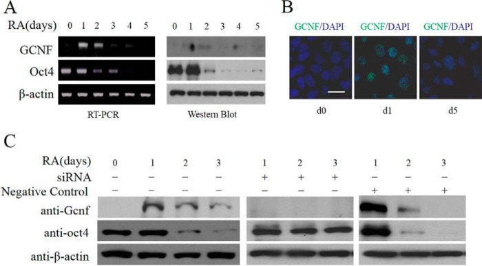
FIGURE 2.
GCNF expression during hES cell differentiation. A, PCR and Western blot results of GCNF and Oct4 expression in undifferentiated and differentiated hES cells. B, immunostaining of GCNF in undifferentiated hES cells (d0) and differentiated hES cells (d1 and d5) treated with RA. C, siRNA-mediated inhibition of GCNF during hES cell differentiation; Western blot results of GCNF and Oct4 expression; β-actin was used as a loading control. Left panel: samples without treatment; middle panel: samples with GCNF siRNA treatment; right panel: negative control (NC) with non-targeting siRNA treatment. Scale bar: 20 μm.

