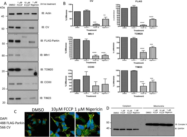
FIGURE 6.
24-h treatment of cells induced similar effects of FCCP and nigericin on mitophagy. A and B, FLAG-Parkin SH-SY5Y neuroblastoma cells were treated with either 1 μm nigericin, 1 μm FCCP, 10 μm FCCP, or DMSO control for 24 h prior to protein extraction. Whole cell lysates were separated on SDS-PAGE gels and Western blotted for mitochondria (CV, TOM20, COXII, TIM23) and mitophagy pathway (FLAG-Parkin and mitofusin) markers, while actin is used as a loading control. The same protein loading was used for each lane in the Western blots. Representative Western blots from n = 3 independent experiments. Significance was determined using Anova (Dunnetts Multiple comparisons) **, p = 0.01. C, immunocytochemistry images show a diffuse Parkin (anti-FLAG, green) pattern and a network of mitochondria (anti-CV, red) in control cells. Upon incubation of 10 μm FCCP or 1 μm nigericin for 24 h, Parkin (FLAG) is recruited to mitochondria (CV) indicated by yellow co-localization. D, FLAG-Parkin SHSY5Y neuroblastoma cells were treated with either 1 μm nigericin, 1 μm, 10 μm FCCP, or DMSO control for 2 h prior to cell lysate fractionation (mitochondria enrichment) and SDS-PAGE. Fractionation efficiency was determined by Western blotting cytoplasmic and mitochondrial-enriched samples using CV (mitochondrial) and GAPDH (cytosolic).

