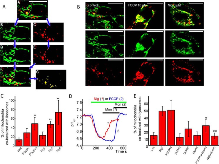
FIGURE 7.
pH-induced changes in co-localization of mitochondria and lysosomes. A, mitochondria to lysosomes colocalization. Double channel confocal image (A) was split into green (mitochondria, Ai) and red (lysosomes, Aii) channels using ImageJ software. Both 8-bit images were thresholded (Aiii, Aiv). Mitochondrial areas were calculated. Areas where mitochondria and lysosomes overlap (yellow) were identified (Av, Avi), and their area was calculated. B, images of experiments with a 2-h pretreatment of the SH-SY5Y neuroblastoma cells with 10 μm FCCP or 6 μm nigericin increase the number of mitochondria (MitoTracker Green, green) localized in lysosomes (LysoTracker red, red). The bar is 20 μm. C, percentage of mitochondria localized in lysosomes after 2 h of pretreatment with different concentrations of FCCP and nigericin. D, effect of monensin on FCCP and nigericin-induced acidification of cytosol of SH-SY5Y neuroblastoma cells. E, percentage of mitochondria localized in lysosomes pretreated with 3 μm nigericin or 10 μm FCCP for 30 min with or without subsequent treatment of the cells with 10 μm monensin. Monensin (10 μm) and 10 or 50 μm DMA were also tested without FCCP or nigericin.

