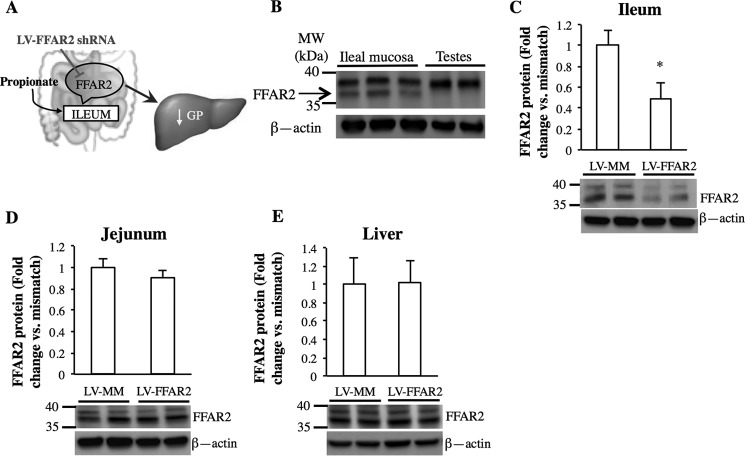
FIGURE 2.
Expression of FFAR2 is reduced in the ileal mucosa after infection of LV-FFAR2 shRNA. A, schematic representation of the working hypothesis. B, Western blot of the presence of FFAR2 in the ileal mucosa of rats. C–E, representative Western blots and quantitative analysis of FFAR2 protein expression normalized to β-actin in the ileum (C), jejunum (D), and liver (E) of rats with ileal infection of either LV-FFAR2 shRNA or LV-MM (*, p < 0.05; calculated by unpaired t test; n = 4 for each).

