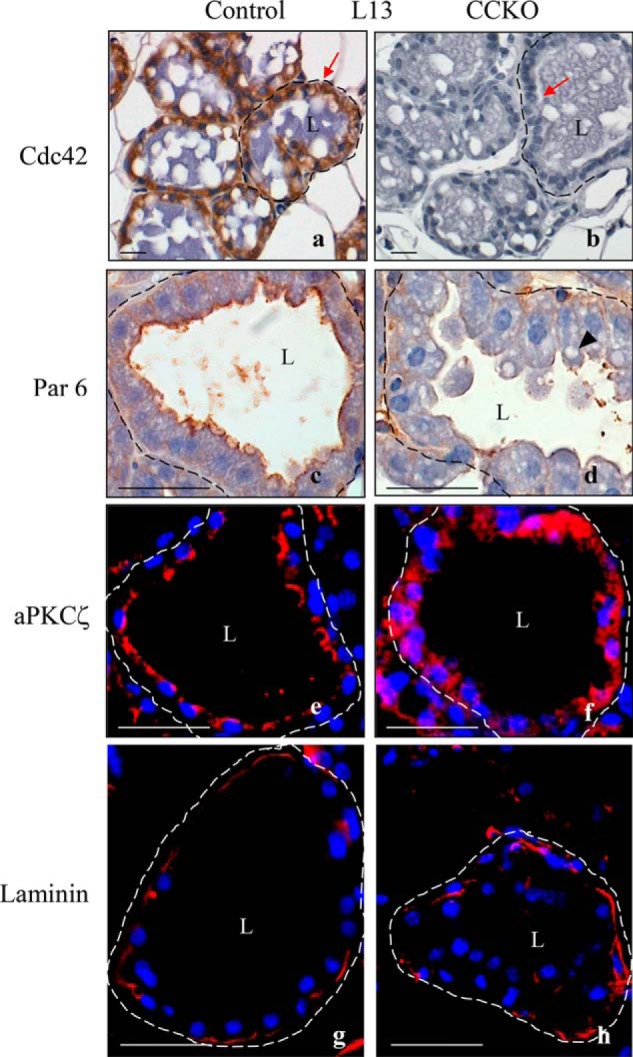
FIGURE 4.

Cdc42 deletion disrupts apical/basal polarity in mammary alveolar epithelial cells. a, b, immunohistochemistry images of 5 μm-thick sections from lactation day 13 depicting Cdc42 expression (brown) in alveolar luminal epithelial cells of the mammary gland from a control mouse (red arrow in a), as compared with a CCKO mouse (red arrow in b). c, d: immunohistochemistry images depicting the localization of Par6 at lactation day 13. Arrowhead in d indicates lipid accumulations. e and f, immunofluorescence images showing the localization of aPKCζ (red) at lactation day 13. Nuclei are stained blue. g and h, immunofluorescence images depicting laminin deposition (red) surrounding an alveolus from control and CCKO mammary glands. Nuclei are stained blue. Dashed lines outline representative alveoli. L marks the central lumen of an alveolus. Scale bars represent 50 μm. The results are representative of the analysis of >3 mice each from control and CCKO groups.
