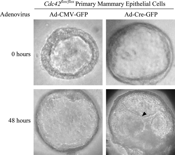
FIGURE 6.
Cdc42 deletion causes luminal filling in primary mammary epithelial cells. Phase contrast images depicting primary mammary epithelial cells forming acini with hollowed lumens at the time of adenovirus transduction (top panels). Forty-eight hours after transduction, control (Ad-CMV-GFP) acini retain hollowed lumens (bottom left panel), while Cdc42-deletion (Ad-Cre-GFP) acini display cells that have migrated into the lumen (arrowhead, bottom right panel).

