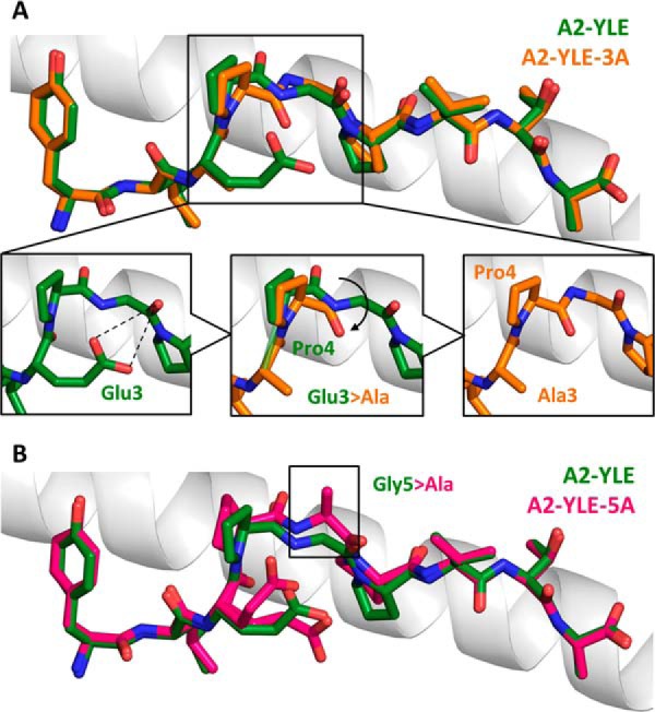
FIGURE 5.

Conformational comparison of YLE, YLE-3A, and A2-YLE-5A peptides presented by HLA-A*0201. A, YLE (dark green sticks) and YLE-3A (orange sticks) peptide alignment by superimposition of HLA-A*0201 α1 helix (gray schematic). Boxed residues indicate the mutation of Glu3 into an alanine. The insets show how the Glu3 → Ala substitution causes a shift in position (black arrow) of neighbor residue Pro4 in the A2-YLE-3A structure compared with the A2-YLE structure. B, YLE (dark green sticks) and YLE-5A (pink sticks) peptide alignment by superimposition of HLA-A*0201 α1 helix (gray schematic). Boxed residues indicate the mutation of glycine 5 into an alanine.
