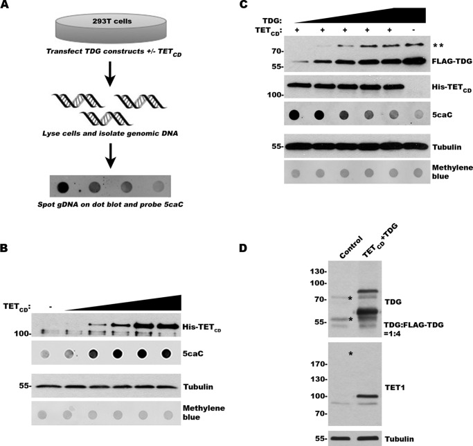
FIGURE 1.
TETCD expression and induction of 5caC can be used to assess TDG activity in vivo. A, schematic of the experimental approach to evaluate TDG activity in vivo. B, expression of TETCD induces formation of 5caC in a dose-dependent manner. The cells were transfected with increasing levels of TETCD expression vector. Protein expression and 5caC levels were monitored by immunoblot analysis. Protein and DNA loading were assessed by tubulin and methylene blue staining, respectively. C, expression of exogenous TDG suppresses 5caC accumulation in a dose-dependent manner. The cells were transfected with TETCD and varying levels of TDG. Protein expression and 5caC levels were monitored as in B. ** indicates sumoylated TDG. D, immunoblot analysis of control transfected cells and cells transfected with TETCD and FLAG-TDG. The blots were probed with TET1- and TDG-specific antibodies for detection of both endogenous and exogenous proteins. Asterisks indicate the positions of endogenous SUMO-TDG, TDG, and the predicted size of endogenous TET1 (not detected). The ratio of TDG to FLAG-TDG was determined using ImageJ.

