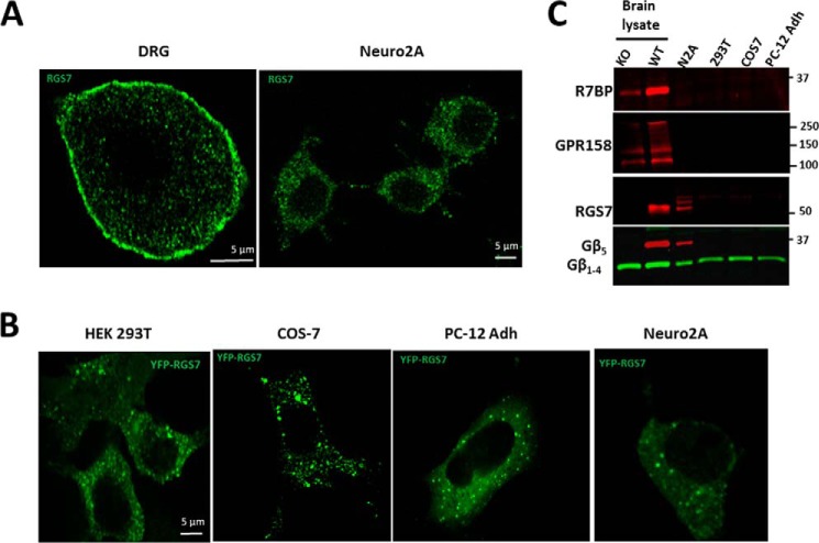
FIGURE 1.
Subcellular localization of endogenous and overexpressed RGS7. A, adult mouse day 6 in vitro DRG neurons (left panel) or N2A cells (right panel) were stained with anti-RGS7 antibodies as described under “Experimental Procedures.” B, indicated cell lines were transfected with plasmids expressing YFP-RGS7 and Gβ5, and YFP was directly imaged by fluorescence microscopy. C, lysates from indicated cell lines were analyzed by Western blot for R7BP, GPR158, RGS7, Gβ5, and G protein β subunit (1–4) expression. Wild-type and Gβ5 knock-out mouse brains were used as a control. Shown are representative immunoblots from two experiments.

