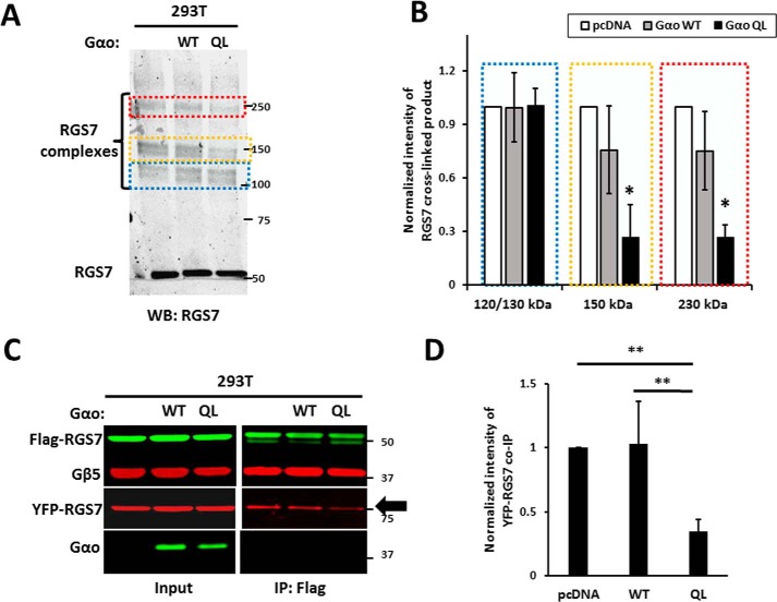
FIGURE 9.
Effect of Gαo subunit on RGS7 homo-oligomerization. A, HEK293T cells transfected with RGS7, Gβ5 with or without wild-type Gαo or its constitutively active Q205L (QL) mutant. The ratio of plasmid DNA in transfection was 4:1:5 (RGS7, Gβ5, Gαo, or pcDNA, respectively). After PFA-induced cross-linking, the cell lysates were analyzed by immunoblot using RGS7 antibody. The areas denoted by the colored broken lines highlight the positions of the 120/130-kDa Gβ5-RGS7 conjugate, the 150-kDa RGS7, and 250-kDa Gβ5-RGS7 oligomers. WB, Western blot. B, quantification of the data in A. The normalized band intensity in the control (pcDNA) was set to 1.0. Data are means ± S.D., n = 3. *, p <0.05. C, lysates from cells expressing FLAG-RGS7, YFP-RGS7, Gβ5, with or without wild-type or mutant Gαo were subjected to immunoprecipitation using FLAG antibody. The eluates were analyzed by immunoblot using FLAG, YFP, Gβ5, and Gαo antibodies. D, quantification of data shown in C. YFP-RGS7 co-IP intensity was normalized to FLAG-RGS7 IP. The results are expressed as means ± S.D.; **, p <0.01.

