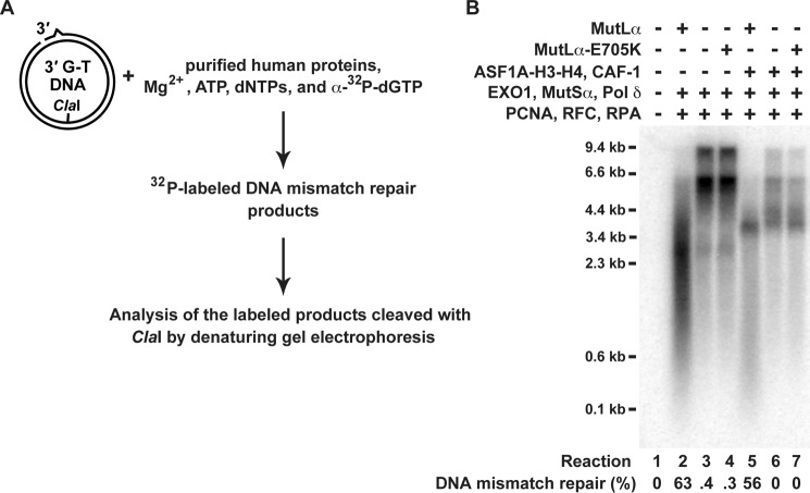
FIGURE 7.
Analysis of DNA molecules labeled with [32P]dGMP during the course of the reconstituted excision-dependent MMR reactions. The reaction mixtures included 33 μCi/ml [α-32P]dGTP (3000 Ci/mmol). The other reaction conditions are described in Fig. 6. A fraction of each recovered DNA was cleaved with ClaI and HindIII to score MMR, and the rest was cleaved with ClaI and separated on a denaturing agarose gel. The gel was dried and exposed to a phosphorimaging screen. The image was generated with a Typhoon biomolecular imager (GE Healthcare). A, outline of the experiment. B, [32P]dGMP-labeled DNA molecules formed in the indicated reactions. The level of MMR in each of the reactions is also shown. The MMR data are the averages ± 1 S.D. (n ≥ 2).

