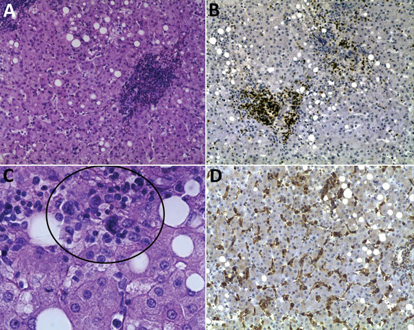
Figure 2.

Histopathologic appearance of liver biopsy sample from woman with fatal human monocytic ehrlichiosis, Mexico, 2013. A) Clusters of cells in the liver lobule. Hematoxylin and eosin (H&E) stain; original magnification ×200. B) Immunohistochemical detection of T lymphocytes (CD3). Original magnification ×100. C) Multinucleated cells in parenchyma (circle). H&E stain; original magnification ×400. D) Immunohistochemical detection of macrophages and hyperplasia of Kupffer cells (CD68). Original magnification ×100.
