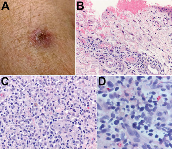
Figure 2.

A) Eschar on the right arm of patient 1 at the site of tick bite sustained in Santa Cruz County, Arizona, USA. B) Histological appearance of the eschar biopsy specimen showing ulcerated epidermis with hemorrhage and perivascular lymphohistiocytic inflammatory infiltrates in the superficial dermis. Hematoxylin-eosin staining; original magnification ×50. C) Dense lymphohistiocytic infiltrates around eccrine ducts in the deep dermis of the biopsy specimen. Hematoxylin-eosin staining; original magnification ×100. D) Sparsely distributed intracellular antigens of Rickettsia parkeri (red) within the inflammatory infiltrates, detected by immunohistochemistry. Alkaline phosphatase with naphthol-fast red and hematoxylin counterstaining; original magnification ×158.
