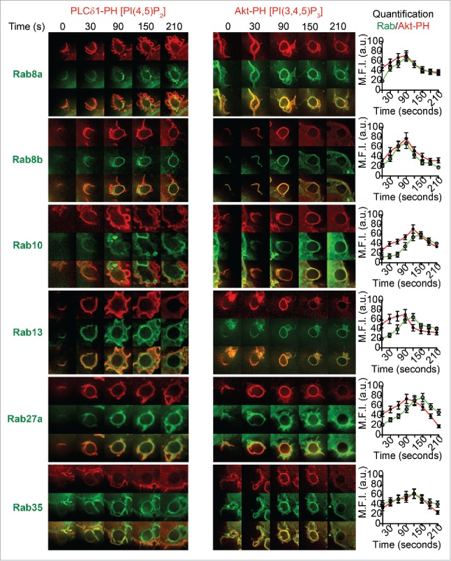
Figure 3.

Spatiotemporal recruitment of Rabs during enrichment of PI(4,5)P2 and PI(3,4,5)3 at the phagocytic cups in live macrophages. RAW 264.7 macrophages were cotransfected to express GFP-tagged Rab8a, Rab8b, Rab10, Rab13, Rab27a, or Rab35 and either mCherry-PLC-PH (A) or mCherry-Akt-PH (B). Representative time lapse images are shown from 0 s to 210 s where 0 s represents the IgG-sRBC making contact with the cell surface. The quantification of maximum fluorescence intensities from GFP-Rabs and mCherry-Akt-PH are shown on the right, revealing the enrichment levels of these proteins throughout phagocytosis. Data are shown as mean ± SEM, n = 6 phagosomes from 3 different cells.
