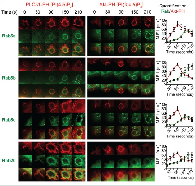
Figure 6.

Spatiotemporal recruitment of Rabs after enrichment of PI(4,5)P2 and PI(3,4,5)3 on phagosomes in live macrophages. RAW 264.7 macrophages were cotransfected to express GFP-tagged Rab5a, Rab5b, Rab5c, or Rab20 and either mCherry-PLC-PH (left panels) or mCherry-Akt-PH (right panels). Representative time lapse images of each sample are shown from 0 s to 210 s where 0 s represents the first instance where the plasma membrane contacts with the IgG-sRBC, detected by enrichment of mCherry-PLC-PH/Akt-PH at the base of the phagocytic cup. Quantification of fluorescence intensities for GFP-Rab in relation to mCherryAkt-PH throughout phagocytosis is shown in the graphs on the right. Fluorescence is plotted using the mean fluorescence intensity around the circumference of the IgG-sRBC from the GFP and mCherry channels and data are shown as mean ± SEM, n = 6 phagosomes from 3 different cells.
