. 2016 May 9;5:e13998. doi: 10.7554/eLife.13998

Figure 4. Regenerated neurons show electrophysiological features similar to uninjured pallium neurons and receive afferent input.

(a) Schematic representation of whole-cell patch clamp recording and subsequent biocytin filling. (b) Representative images of EdU+/Biocytin+ neurons in the injured pallium. (c) Representative traces of the membrane voltage changes in response to depolarizing and hyperpolarizing current steps injections of neurons from uninjured and injured animals. (d) Zoom-in of single action potential evoked by a depolarizing current step recorded in neurons of dorsal pallium. (e) Summary of passive and active electrophysiological properties: membrane capacitance (Cm), membrane resistance (Rm), resting membrane potential (RMP), action potential (AP) threshold, AP height and AP half width. (f) Sample traces of spontaneous postsynaptic currents (sPSCs) recorded under voltage clamp (Vh = −70 mV). (g) Summary of sPSCs features in neurons from uninjured and injured animals. All results are expressed as the mean ± SD. *p<0.05; unpaired, two-tailed Student’s t-test.

DOI: http://dx.doi.org/10.7554/eLife.13998.013

Figure 4.

Figure 4—figure supplement 1. EdU+ neurons in the injured pallium show immature electrophysiological properties.

Figure 4—figure supplement 1.

(a) Changes of the membrane voltage in response to depolarizing and hyperpolarizing current steps injections from an EdU+ neuron recorded in the injured pallium. (b) Zoom-in of single action potential evoked by a depolarizing current step. Broad and short action potential is typical of immature neurons. (c) Sample trace of spontaneous postsynaptic currents (sPSCs) recorded under voltage clamp (Vh=−70 mV).
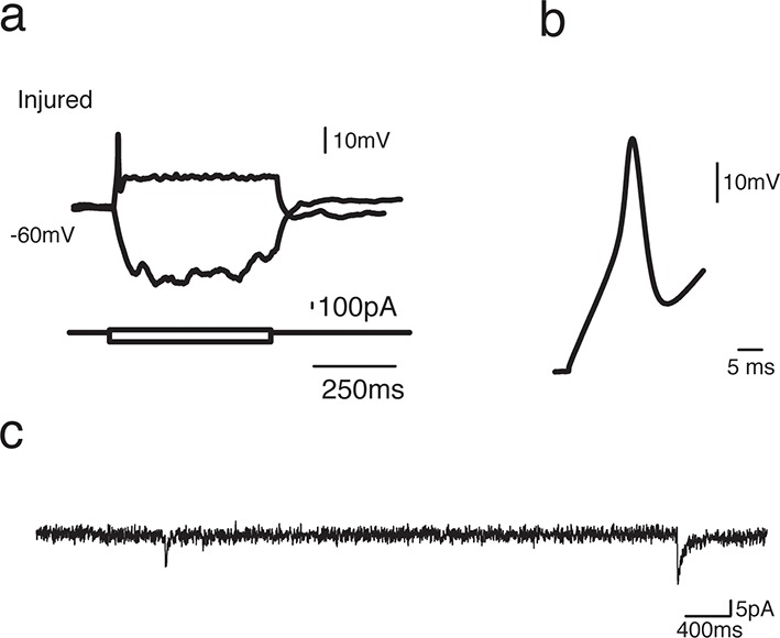
Figure 4—figure supplement 2. Most sPSCs are abolished by administration of AMPA receptor blocker.

Figure 4—figure supplement 2.

(a) Sample trace of spontaneous postsynaptic currents (sPSCs) recorded under voltage clamp (Vh=−70 mV). (b) Sample trace of the same neuron after application of AMPA receptor blocker, NBQX [10 µM], which abolishes most sPSCs.
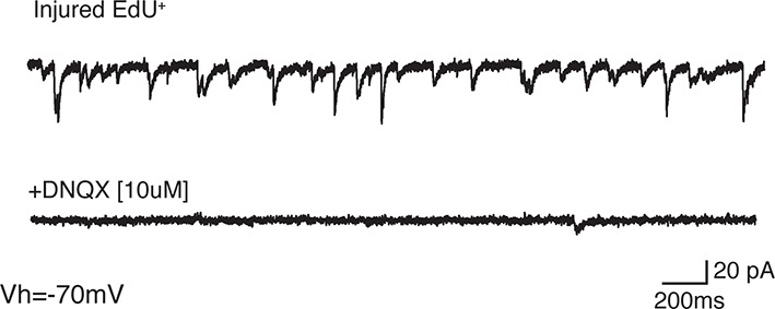
Figure 4—figure supplement 3. Spontaneous calcium transients in neurons do not differ between injured and control dorsal pallium.

Figure 4—figure supplement 3.

(a) Schematic of ex vivo calcium imaging. (b) Representative image of Fluo-4 staining in the dorsal pallium after dye loading (left panel). Region of interest (ROI) segmentation for individual neurons (middle panel) and merge (right panel). (c) Representative traces of spontaneously generated calcium transients in labeled neurons within dorsal pallium. (d) Raster plot of peaks in traces from panel c. (e) Quantification of the peak amplitude (left panel), rise time (middle panel), and fall time (right panel) in individual neurons shows no significant difference between injured and control dorsal pallium. V, ventricle. All results are expressed as the mean ± SEM. Unpaired, two-tailed Student’s t-test.