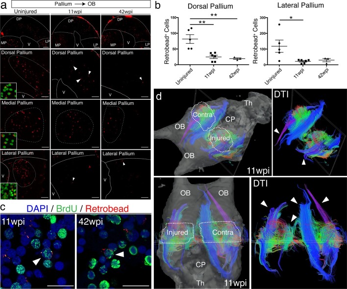
Figure 6. Long-distance projections from the regenerated dorsal pallium are significantly reduced.
(a) Retrograde tracing from the olfactory bulb shows reduced number of labeled cells in the dorsal pallium and lateral pallium at 11wpi (middle panels) and 42wpi (right panels) compared to uninjured control (left panels). Insets, high-magnification images of DAPI (green) and Retrobeads (red). (b) Quantification of the number of Retrobead+ cells in the dorsal and lateral pallium at different time points after injury. (c) Representative immunohistochemistry images of BrdU+/Retrobead+ cells in the regenerated dorsal pallium at 11wpi (left panel) and 42wpi (right panel). (d) ex vivo DTI of 11wpi brains show disruption of fiber tracts (arrowheads) in the injured hemisphere. White dotted lines (left panels) represent regions of interest in injured and contralateral hemispheres. OB, Olfactory Bulb; CP, Choroid Plexus; Th, Thalamus; V, Ventricle; DP, Dorsal Pallium; MP, Medial Pallium; LP, Lateral Pallium. Scale Bars; 50 μm (b), 20 μm (d). All results are expressed as the mean ± SEM. *p<0.05, **p<0.01; one-way ANOVA with post-hoc Tukey’s multiple comparison test.


