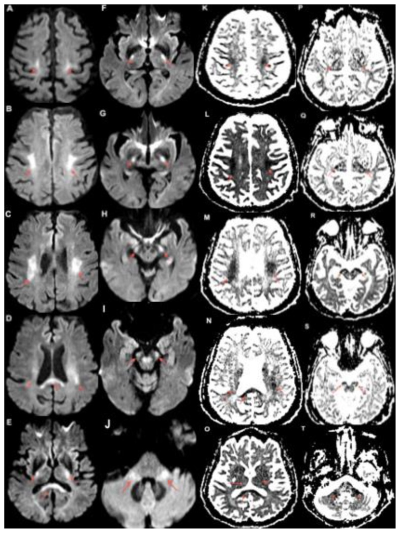
Figure 1.
80 year old female with capecitabine-induced leukoencephalopathy.
FINDINGS: Diffusion weighted MRI imaging demonstrates restricted diffusion along the course of the bilateral corticospinal tracts. The bilateral centrum semiovale (A, B, K, L), corona radiata (C, D, M, N), posterior limbs of the internal capsules (E, F, G, P, Q, R) and cerebral peduncles (H, I, R, S) are involved. There is also involvement of the corpus callosum (D, E, N, O) and middle cerebellar peduncles (J, T) seen.
TECHNIQUE: MRI. Magnet Strength: 3.0 Telsa. Plane: Axial. DWI b=1000 s/mm2 TE: 101ms TR: 4700ms FOV: 230mm × 230mm. Matrix: 192 × 192 Slice Thickness: 4.00mm

