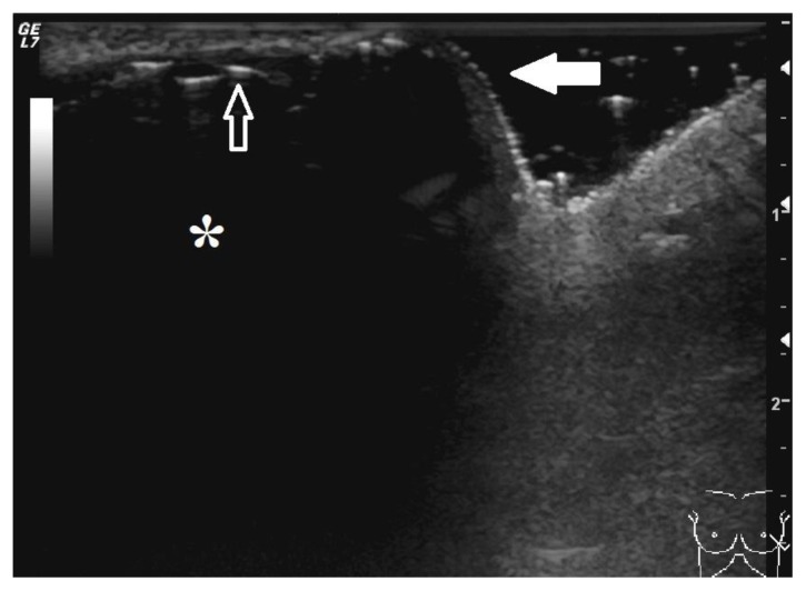
Figure 5.
44-year-old male with left pectoral implant displacement.
FINDINGS: Sonographic image of the displaced implant at site of impending extrusion. The implant (*) is anechoic. The thin echogenic line (open arrow) that is present between the anechoic implant and skin represents the anterior margin of the implant. The posterior margin of the implant is not visualized due to attenuation of the ultrasound beam by the silicone. The overlying pectoralis muscle is not readily appreciated and the implant is directly subjacent to the skin (closed arrow).
TECHNIQUE: Ultrasound, radial plane, 14 megahertz transducer.

