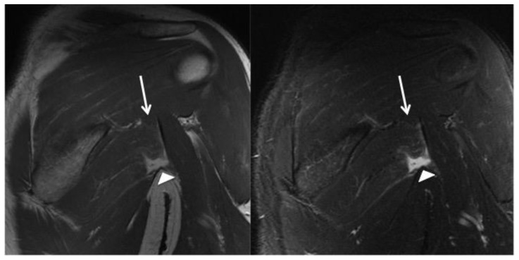
Figure 2.
53 year-old man with isolated left teres major rupture.
FINDINGS: Normal location of the teres major inferior to the scapula. There is partial (grade II myotendinous junction) tearing of the superior (cranial) most fibers, which demonstrate mild increased signal intensity and an undulating contour (arrows). There is a complete tear (grade III myotendinous injury) of the inferior (caudal) tendon fibers with a fluid filled gap at the site of the complete tear (arrowheads).
TECHNIQUE: Coronal oblique intermediate fast spin echo [TR: 3500 TE: 37.128; field of view 24 cm; matrix 384 × 288; slice thickness: 4 mm with no gap] (left) and short-tau inversion recovery [TR: 2750; TE: 50.48; field of view: 24; matrix: 288 × 224; slice thickness 5mm with no gap] (right).

