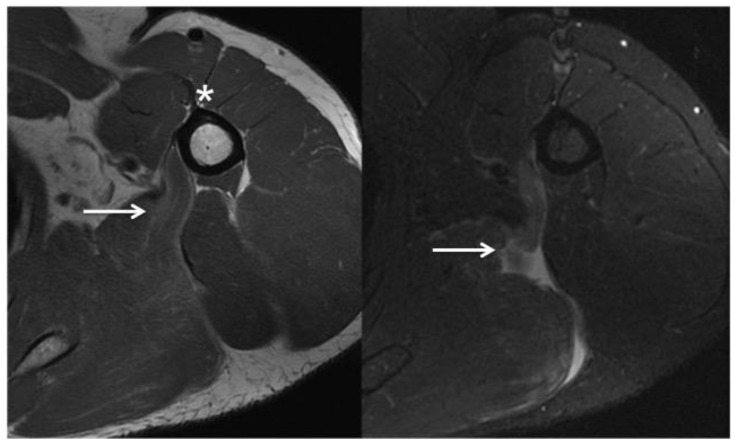
Figure 3.
53 year-old man with isolated left teres major rupture.
FINDINGS: The teres major demonstrates tapering of the muscle fibers from posterior medial to anterior lateral. The normal teres major tendon attaches to the medial ridge of the intertubercular groove, just posterior medial to the pectoralis major tendon (asterisk, left). Intermediate fast spin echo images of the superior tendon fibers demonstrate mild increased signal intensity and undulation of the fibers compatible with partial tearing (arrow, left). T2-weighted fat saturated images more inferiorly demonstrate complete tearing of the inferior-most muscle fibers with a fluid filled gap at the site of tearing (arrow, right).
TECHNIQUE: Axial intermediate fast spin echo [TR: 3850 TE: 33.408; field of view 22cm; matrix: 384 × 288; slice thickness: 4 mm with no gap] (left) and short-tau inversion recovery [TR: 3,500; TE: 49.28; field of view: 22; matrix: 288 × 224; slice thickness 5mm with no gap] (right).

