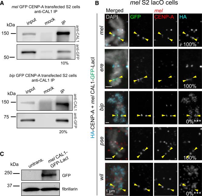
Figure 2. D. melanogaster CAL1 Cannot Recruit D. bipectinata CENP-A to an Ectopic Locus.
(A) Western blots of IPs with anti-CAL1 antibodies from nuclear extracts transiently expressing mel GFP-CENP-A (top) or bip GFP-CENP-A (bottom). IP was confirmed using anti-CAL1 antibody (top blot). Presence of GFP-CENP-A in CAL1 pull-downs was detected with anti-GFP antibody (bottom blot). Shown is the percentage of immunoprecipitated GFP-CENP-A relative to input.
(B) Representative IF images of metaphase chromosome spreads from mel S2 lacO cells transiently co-expressing mel CAL1-GFP-LacI and HA-CENP-A orthologs: mel (top); ere (second); bip (third); pse (fourth); and wil (bottom). Chromosome spreads were quantified for the presence of HA-CENP-A at the lacO site (percentage shown in right column). Endogenous mel CENP-A is shown in red, HA in aqua, GFP in green, and DAPI in gray. Note that mel CENP-A antibodies are specific for this species and that upon expression of CENP-A orthologs that localize to mel centromeres, endogenous CENP-A levels decrease (e.g., ere CENP-A; see also Figure S2). This is not observed for mel HACENP-A, as mel CENP-A antibodies recognize this tagged protein. ***p < 0.0001 (Fisher's two-tailed test) for bip or wil CENP-A compared with mel CENP-A recruitment at the lacO. n = 13 spreads for mel CENP-A recruitment, 8 for ere, 11 for bip, 15 for pse, and 8 for wil. Yellow arrowheads indicate the lacO array. These results were confirmed by one biological replicate with the CAL1-GFP-LacI construct, and two biological replicates with GFP-CENP-A and CAL1-LacI constructs (data not shown).
(C) Western blots with anti-GFP (top) and anti-fibrillarin (loading control, bottom) antibodies of whole-cell extracts showing the expression of induced mel CAL1-GFP-LacI in lacO cells shown in (B).

