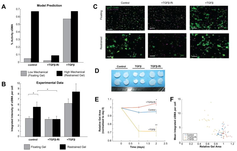
Fig. 7. Experimental validation of predicted role for TGFβ in mechanical-induced expression of αSMA.
(A) The model predicted that high mechanical input would increase αSMA expression, but that this effect would be mitigated by a TGFβ-R inhibitor. TGFβ increased αSMA at both low and high mechanical input. (B) Experimental measurements of αSMA expression as measured by immunofluorescence in adult rat cardiac fibroblasts cultured in floating or restrained gels, in which fibroblasts experienced increased mechanical stimulation. As predicted by the model, restrained gels exhibited increased αSMA expression, which was mitigated by the TGFβ-R inhibitor (TGFβ-Ri) SD208. (C) Example images of cardiac fibroblasts cultured in restrained and floating gels, stained for αSMA (green) and DAPI (purple). Scale bar = 100 μm. (D) Increased compaction of floating gels treated with TGFβ but not with TGFβ-Ri. (E) Floating gels compact over time with TGFβ and become more relaxed over time after treatment with TGFβ-Ri. (F) The final size of floating gels was inversely correlated with αSMA expression. * indicates p<0.05, and ** indicates p<0.01. All error bars indicate standard error of the mean.

