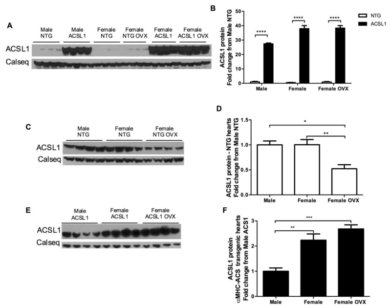
Figure 5. Cardiac ACSL1 protein expression analysis.
(A and B) Western blot results displaying elevated ACSL1 content ACSL1 hearts versus NTG (n = 4 all). (C and D) Western blot results for ACSL1 content in NTG hearts. ACSL1 protein is similar between NTG males and female hearts, but decreases with ovariectomy (OVX). Female sex hormones are necessary for normal female ACSL1 expression. (E and F) Western blot results showing greater overexpression of ACSL1 in female and female OVX hearts versus male hearts. However, the increase in ACSL1 expression compared to respective NTG hearts was similar among sexes (A and B). Loading normalized to calsequestrin (Calseq) as control. *p < 0.05, ** p < 0.01, *** p < 0.001, and **** p < 0.0001.

