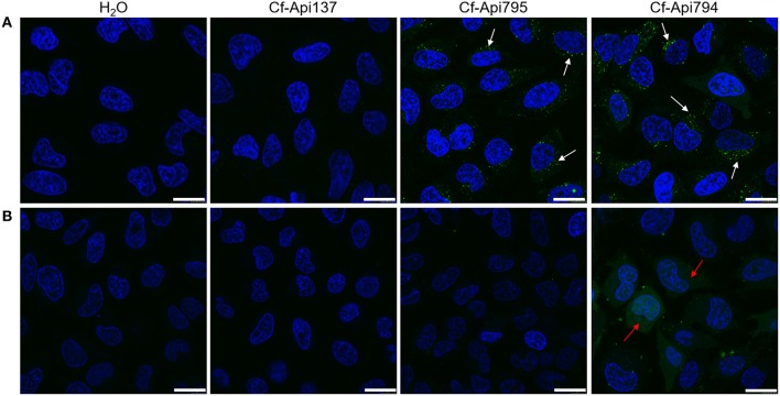
Figure 6.
Confocal microscopy images of HeLa cells incubated with Cf-labeled apidaecin peptides. Cells were treated without (A) or with dynasore (0.2 mmol/L; B) for 45 min prior to peptide treatment (40 μmol/L, 30 min). The cells' nuclei were visualized with Hoechst 33324 (blue). Arrows indicate examples of areas with endosomes (white) and stained cytosol (red). Bars refer to 20 μm.

