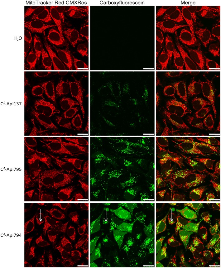
Figure 8.
Localization of the peptides in HeLa cells determined by confocal microscopy. Cells were incubated with the indicated peptide (40 μmol/L) and MitoTracker Red CMXRos (0.1 μmol/L) for 6 h. Bars refer to 20 μm. Arrows mark the slight background fluorescence of the mitochondrial stain and the high fluorescence of the Cf-labeled peptide, which overlap (unspecific colocalization).

