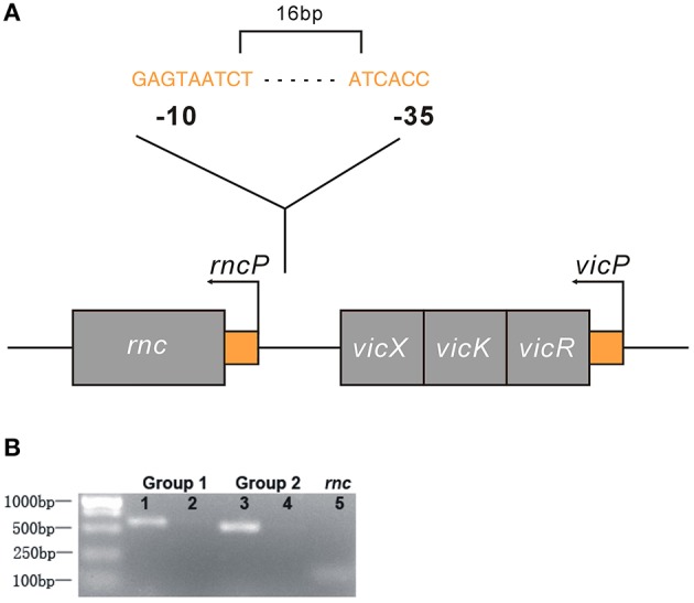
Figure 4.

Genetic analyses of the rnc and vicRKX gene loci. (A) Predicted genetic structure of rnc and vicRKX loci by FGENESB and BPROM. The fragment shown was from the S. mutans chromosome, which contained the intact rnc, vicX, vicK, and vicR genes, shown from left to right. The entire sequence was known for this region. Arrows indicated the initiation sites for the vicP and rncP promoters. The supposed sequences of -10 and -35 boxes in rncP were GAGTAATCT and ATCACC, respectively. (B) PCR of rnc and vicX co-transcription assays. Two sets of specific PCR primers were used for the co-transcription assay. Lanes 1 and 3 were used for UA159 DNA, and lanes 2 and 4 were used for UA159 cDNA, which was reverse transcribed from RNA. Line 5 was used as a reference to verify the feasibility of the cDNA.
