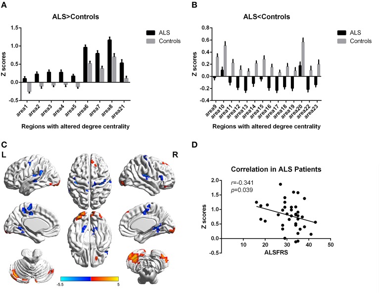
Figure 2.
Regions with altered degree centrality in ALS patients compared with healthy controls in binarized networks. (A,B) represents the regions with increase/decrease degree centrality in ALS patients in bar graphs (group mean Z scores and standard errors of the mean). The red areas in (C) show the regions with increased degree centrality, and the blue areas show the regions with decreased degree centrality. The color bar shows the T values. (D) shows the negative correlation between DC's z-scores in binarized networks of Right inferior Occipital gyrus (area7, BA 18) and the ALSFRS-r scores in ALS group with the age and gender as covariates.

