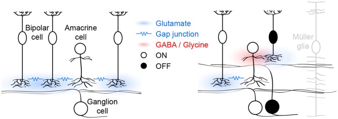
Figure 1.
Schematic of glutamatergic wave circuits. The left panel illustrates the lateral excitatory (blue) network of ON cone bipolar and amacrine cells, which generates and propagates stage III waves. Activity spreads by lateral glutamatergic transmission and gap junctions among neighboring cells. The right panel illustrates the vertical inhibitory (red) pathway through which ON cone bipolar cells hyperpolarize OFF cone bipolar cells and delay excitatory input to and spiking off OFF ganglion cells. Müller glia limit the spread of glutamate and maintain temporal separation of ON and OFF activity.

