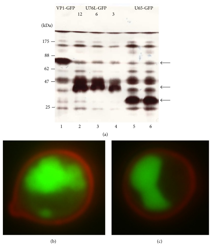
Figure 5.
(a) SDS-PAGE of proteins from cells expressing the following: Lane 1, VP1-GFP (70 kDa, arrow); Lanes 2–4, U76L-GFP (36.2 kDa, arrow, loads of 12, 6, and 3 μL, resp.); Lanes 5 and 6, U65-GFP (34.1 kDa, arrow, independent cultures). (b) Fluorescence microscopy of cells expressing U65-GFP. Cell wall glucan is stained by Congo red. (c) pH 11.5 YCPs.

